Erythrocyte Aggregation on:
[Wikipedia]
[Google]
[Amazon]
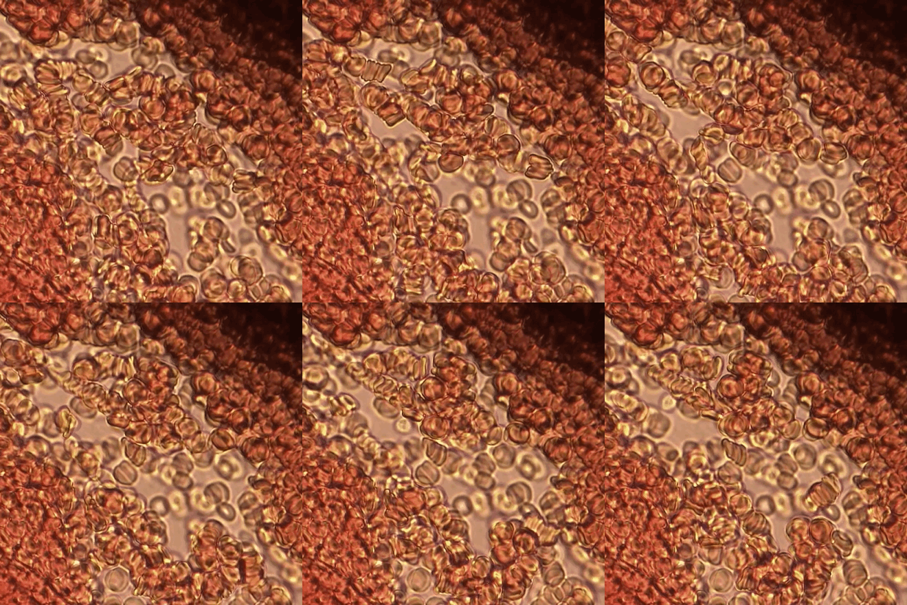
Erythrocyte aggregation is the reversible clumping of red blood cells (RBCs) under low shear forces or at stasis. 

Erythrocyte
Red blood cells (RBCs), also referred to as red cells, red blood corpuscles (in humans or other animals not having nucleus in red blood cells), haematids, erythroid cells or erythrocytes (from Greek ''erythros'' for "red" and ''kytos'' for "holl ...
s aggregate in a special way, forming rouleaux
Rouleaux (singular is rouleau) are stacks or aggregations of red blood cells (RBCs) that form because of the unique discoid shape of the cells in vertebrates. The flat surface of the discoid RBCs gives them a large surface area to make contact wi ...
. Rouleaux are stacks of erythrocytes which form because of the unique discoid shape of the cells in vertebrate
Vertebrates () comprise all animal taxa within the subphylum Vertebrata () ( chordates with backbones), including all mammals, birds, reptiles, amphibians, and fish. Vertebrates represent the overwhelming majority of the phylum Chordata, ...
body. The flat surface of the discoid RBCs give them a large surface area to make contact and stick to each other; thus, forming a rouleau. Rouleaux formation takes place only in suspensions of RBC containing high-molecular, fibrilar proteins or polymers in the suspending medium (often Dextran-2000 in-vitro). The most important protein causing rouleaux formation in plasma is fibrinogen. RBC suspended in simple salt solutions do not form rouleaux.
Mechanism
Erythrocyte aggregation is a physiological phenomenon that takes places in normal blood under low-flow conditions or at stasis. The presence or increased concentrations of acute phase proteins, particularlyfibrinogen
Fibrinogen (factor I) is a glycoprotein complex, produced in the liver, that circulates in the blood of all vertebrates. During tissue and vascular injury, it is converted enzymatically by thrombin to fibrin and then to a fibrin-based blood clo ...
, results in enhanced erythrocyte aggregation.
Current experimental and theoretical evidence supports the mechanism related to the depletion of high-molecular weight molecules (e.g., fibrinogen
Fibrinogen (factor I) is a glycoprotein complex, produced in the liver, that circulates in the blood of all vertebrates. During tissue and vascular injury, it is converted enzymatically by thrombin to fibrin and then to a fibrin-based blood clo ...
) for rouleaux formation. This mechanism is also known as “chemiosmotic hypothesis” for aggregation.
Erythrocyte aggregation is determined by both suspending phase (blood plasma
Blood plasma is a light amber-colored liquid component of blood in which blood cells are absent, but contains proteins and other constituents of whole blood in suspension. It makes up about 55% of the body's total blood volume. It is the intra ...
) and cellular properties. Surface properties of erythrocytes, such as surface charge density strongly influence the extent and time course of aggregation.
Effects
Erythrocyte aggregation is the main determinant of blood viscosity at low shear rate. Rouleaux formation also determinesErythrocyte sedimentation rate
The erythrocyte sedimentation rate (ESR or sed rate) is the rate at which red blood cells in anticoagulated whole blood descend in a standardized tube over a period of one hour. It is a common hematology test, and is a non-specific measure of ...
which is a non-specific indicator of the presence of disease.
Influence of erythrocyte aggregation on in vivo blood flow is still a controversial issue. Enhanced aggregation affects venous hemodynamics. Erythrocyte aggregation also affects hemodynamic mechanisms in microcirculation and vascular control mechanisms.
Causes
Conditions which cause increased rouleaux formation includeinfections
An infection is the invasion of tissue (biology), tissues by pathogens, their multiplication, and the reaction of host (biology), host tissues to the infectious agent and the toxins they produce. An infectious disease, also known as a transmiss ...
, inflammatory and connective tissue
Connective tissue is one of the four primary types of animal tissue, along with epithelial tissue, muscle tissue, and nervous tissue. It develops from the mesenchyme derived from the mesoderm the middle embryonic germ layer. Connective tiss ...
disorders, and cancers
Cancer is a group of diseases involving Cell growth#Disorders, abnormal cell growth with the potential to Invasion (cancer), invade or Metastasis, spread to other parts of the body. These contrast with benign tumors, which do not spread. Poss ...
(most common in multiple myeloma
Multiple myeloma (MM), also known as plasma cell myeloma and simply myeloma, is a cancer of plasma cells, a type of white blood cell that normally produces antibodies. Often, no symptoms are noticed initially. As it progresses, bone pain, an ...
). It also occurs in diabetes mellitus
Diabetes, also known as diabetes mellitus, is a group of metabolic disorders characterized by a high blood sugar level ( hyperglycemia) over a prolonged period of time. Symptoms often include frequent urination, increased thirst and increased ap ...
and is one of the causative factors for microvascular occlusion in diabetic retinopathy
Diabetic retinopathy (also known as diabetic eye disease), is a medical condition in which damage occurs to the retina due to diabetes mellitus. It is a leading cause of blindness in developed countries.
Diabetic retinopathy affects up to 80 perc ...
.
Erythrocyte sedimentation rate closely reflects the extent of aggregation, therefore can be used as a measure of aggregation. Erythrocyte aggregation can also be quantitated by monitoring optical properties of blood during the time course of aggregation process.
Measurement
blood film
A blood smear, peripheral blood smear or blood film is a thin layer of blood smeared on a glass microscope slide and then stained in such a way as to allow the various blood cells to be examined microscopically. Blood smears are examined in the ...
syllectometry
intravital microscopy
high-frequency ultrasound
Preclinical imaging is the visualization of living animals for research purposes, such as drug development. Imaging modalities have long been crucial to the researcher in observing changes, either at the organ, tissue, cell, or molecular level, i ...
Optical coherence tomography
References
{{DEFAULTSORT:Erythrocyte Aggregation Blood cells